- Metastases
- Glioblastoma
- Anaplastic astrocytomas
- Medulloblastoma
- Lymphoma
- Gliosarcoma
- Chordoma
- Chondrosarcoma
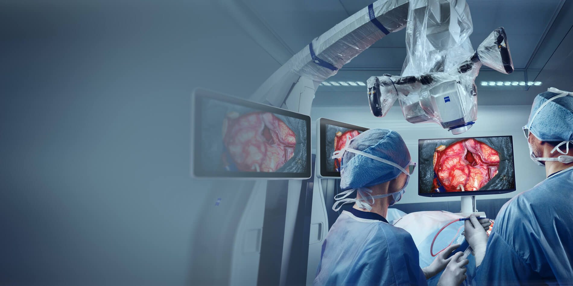
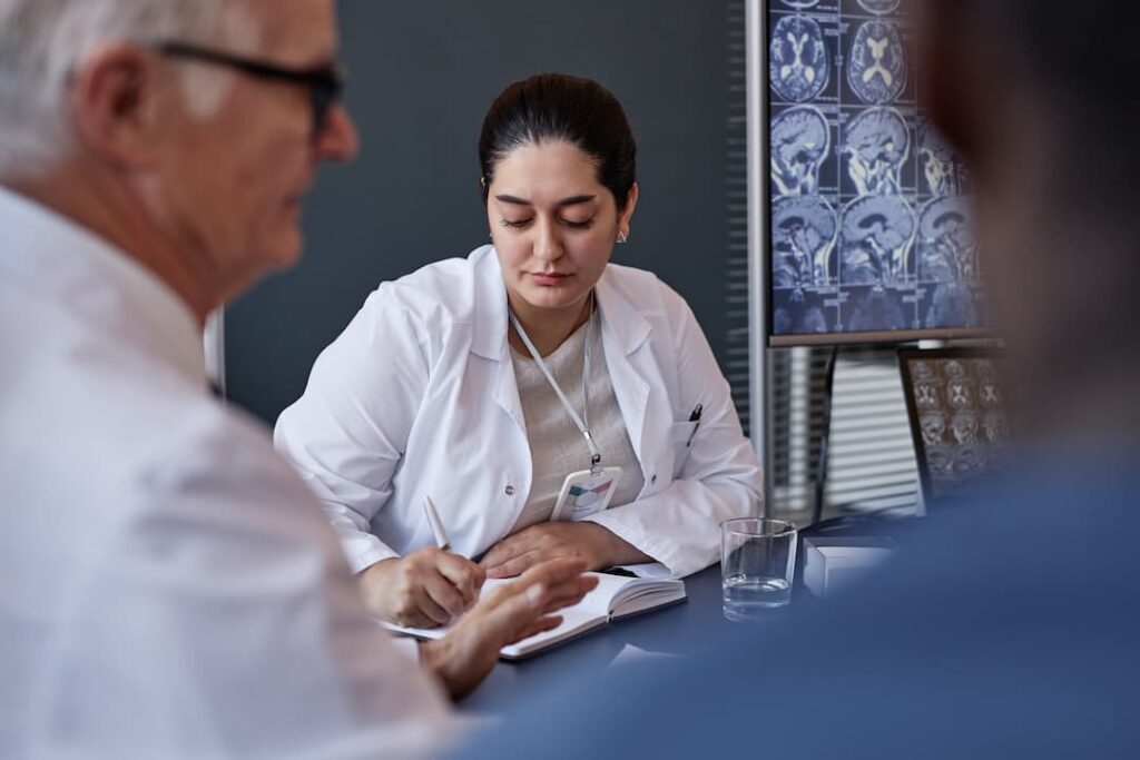
Neuro-oncology institute
Neuro-oncology Institute led by Prof. Gerardo Conesa is a multifunctional medical facility specialized on complex treatment of benign and malignant tumors of the nervous system: brain tumors, spinal tumors and spinal cord tumors.
Experience of our specialists and technical equipment of the hospital allows us
to offer the best treatment for every patient.
Watch the video to know more.
Leading doctors
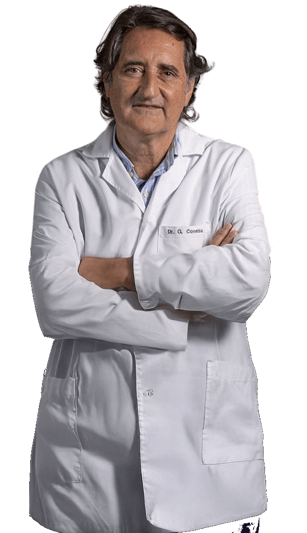
Prof. Gerardo Conesa
Director of Neuro-Oncology Institute, Leading neurosurgeon
Prof. Conesa is the Director of the Institute of Neurosurgery and Neuroscience at Teknon Medical Centre. He earned his medical degree from the University of Barcelona in 1982 and completed his neurosurgery specialization at the University Hospital de Bellvitge in 1990. His training includes internships in both the United States and France, and since 1985 he has served in several university hospitals across Barcelona. He is an active member of numerous professional organizations, including the European Association of Neurosurgical Societies, the European Union of Neurosurgeons, the Spanish Society of Neurosurgery, and the Walter E. Dandy Society.
30+ years of clinical experience
Specialization
- Brain tumors :glioma, glioblastoma, meningioma, neurinoma, brain metastases.
- Lumbar and cervical spine surgery: minimally invasive procedures.
- Epilepsy surgery and Deep Brain Stimulation (DBS).
Treatment
-
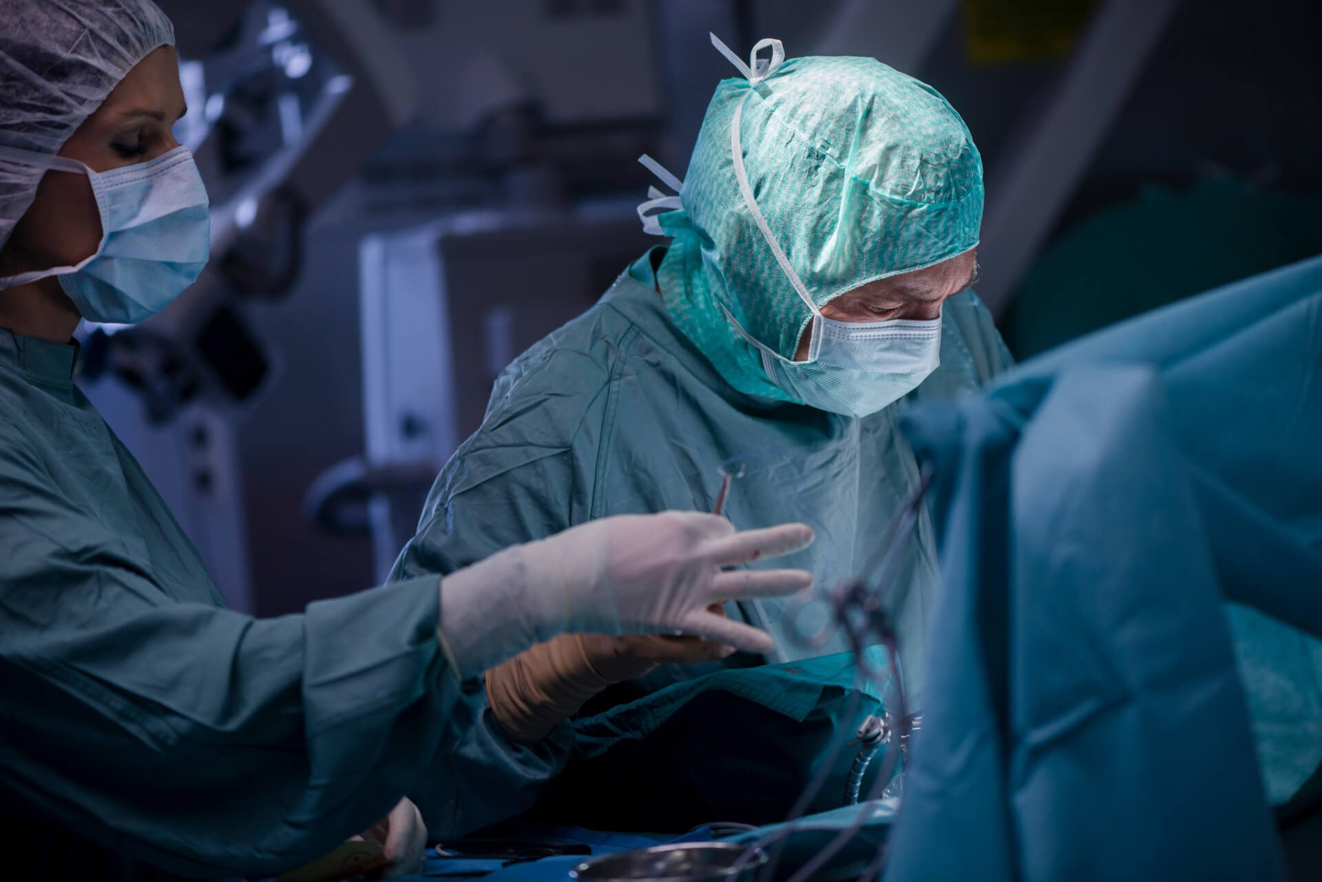
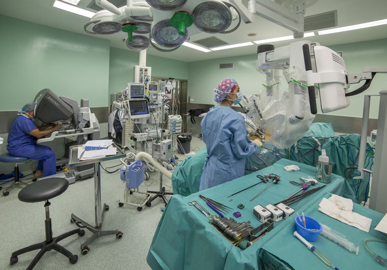
Complete removal or extirpation of the tumor
In almost all cases where the tumor is technically amenable to complete surgical removal, prompt intervention is highly recommended. This action is paramount for mitigating further disease progression and optimizing the patient's overall prognosis.
It must be noted, however, that certain tumor histologies inherently carry a high risk of recurrence even following successful primary extirpation. To consolidate the surgical outcome and prevent relapse, subsequent adjuvant therapy, typically comprising radiation and systemic oncological treatments, is often required.
-
Partial removal or resection of the tumor
When the location of the tumor or its closeness to vital areas makes complete removal unsafe, the medical team may recommend a partial removal (resection).
The main goal of this procedure is maximal safe debulking: removing the largest amount of the tumor possible without causing harm. This is done to significantly reduce the number of cancer cells before other treatments begin, which ultimately improves the patient's long-term outcome and lowers the chance of complications.

Articles
-

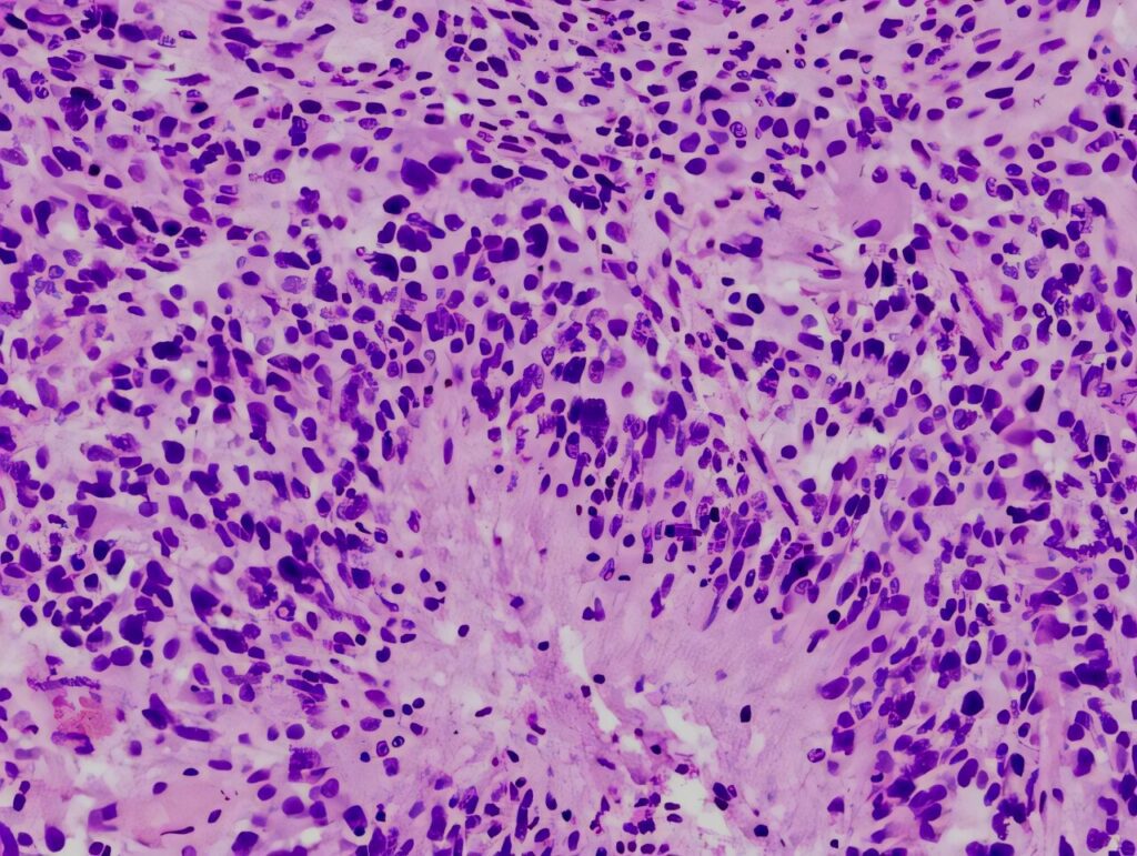
Glioblastoma: Symptoms, Diagnosis, and Treatment
Glioblastoma is the most aggressive primary malignant tumor of the central nervous system in adults. It represents the highest-grade form of diffuse glioma and is characterized by rapid growth, extensive infiltration of surrounding brain tissue, and complex molecular heterogeneity.
Read article -
Meningioma: Symptoms, Diagnosis, and Treatment
Meningiomas are among the most common primary brain tumors, affecting thousands of people each year. Although most meningiomas are benign (non-cancerous), their size and location can still lead to significant neurological problems.
Read article -
Diffuse Astrocytoma: Symptoms, Diagnosis, and Treatment
Diffuse astrocytoma is a primary tumor of the central nervous system arising from astrocytic glial cells. It belongs to the group of infiltrative gliomas and is characterized by slow to moderate growth, diffuse infiltration of surrounding brain tissue, and a variable clinical course.
Read article -
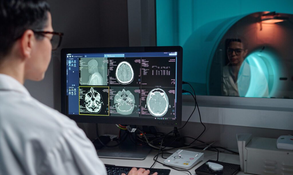
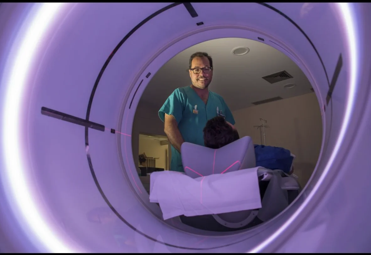
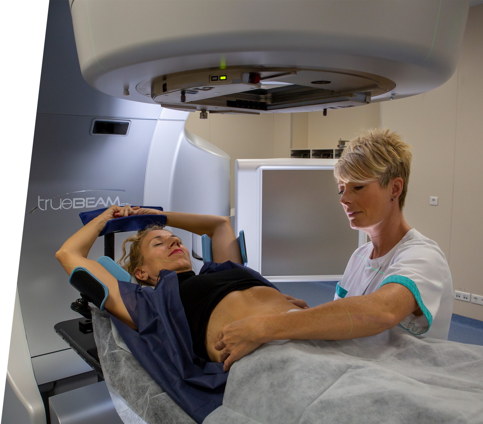
Radiation Therapy in Neurooncology: Principles, Indications, and Long-Term Outcomes
Radiation therapy is a fundamental component of modern neurooncology and plays a central role in the treatment of both primary and metastatic tumors of the central nervous system
Read article
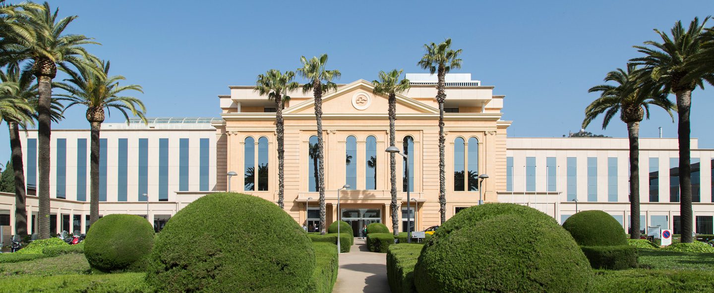
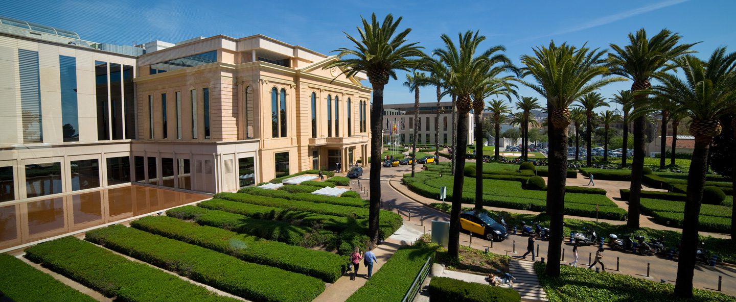
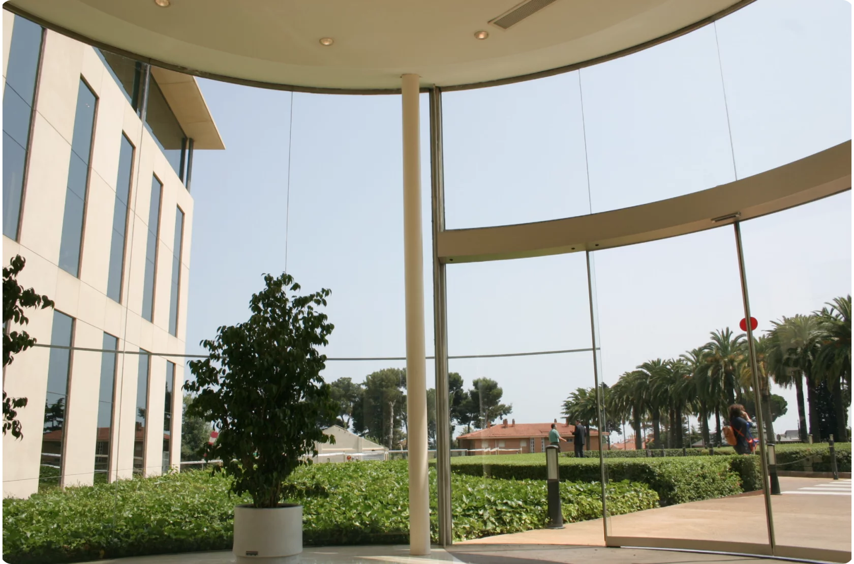
Hospital
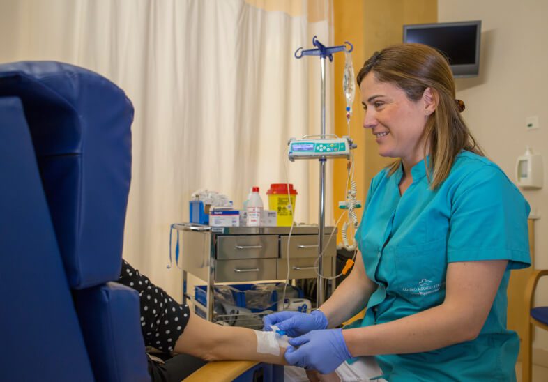
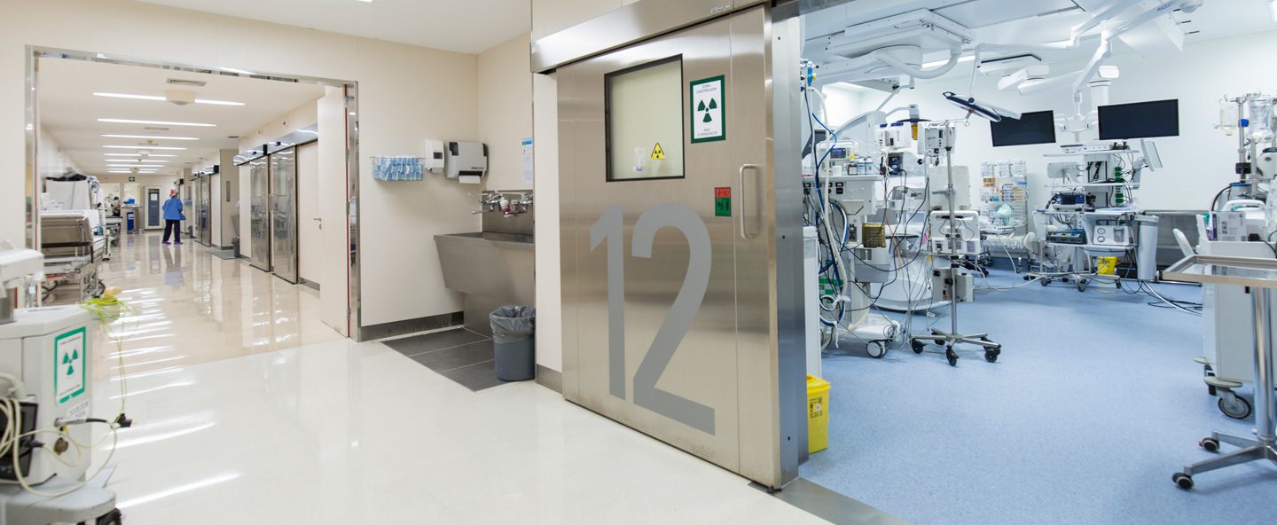
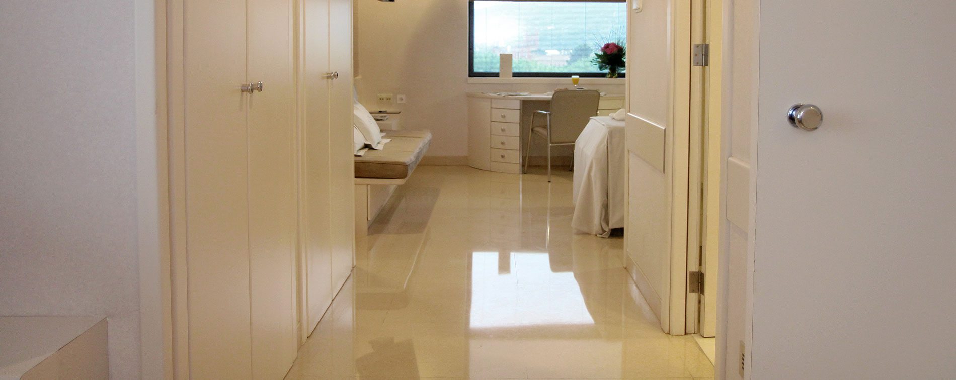
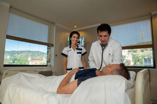
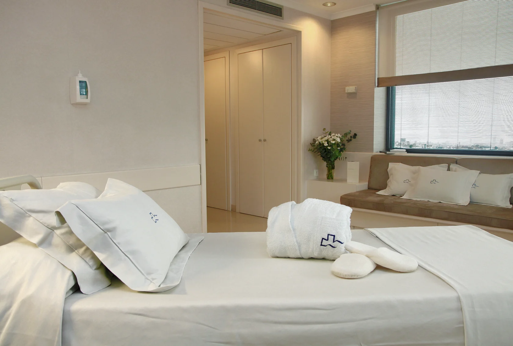
Located at the Teknon Medical Center, one of the largest multidisciplinary hospitals in Spain, the Neuro-oncology Institute guarantees patients access to immediate, comprehensive medical support and expert postoperative care.
Teknon's dedication to quality is confirmed by 8 successful accreditations from Joint Commission International (JCI), ensuring high standards of safety. The hospital's versatility, supported by over 400 doctors and 2,000 specialists, allows for effective management of any potential complications.
This multidisciplinary environment ensures a complete recovery strategy, including individualized diet planning and dedicated physiotherapy.
“Teknon Medical Centre — the best international hospital”
IMTJ: International Medical Travel Journal

For doctors
The Neuro-oncology Institute actively collaborates with physicians and medical centers globally to exchange knowledge and advance shared experience in combating neuro-oncological pathology.
Medical professionals interested in exploring collaborative opportunities are invited to complete the provided form, and we will respond promptly.